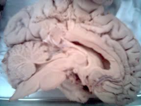
Last week I had the pleasure of visiting Dr. Mark DeSantis’s lab to examine actual preserved human brains with my fellow Research Experience for Undergraduates participants. Dr. DeSantis was kind enough to provide a tour of the structures and functions of some of the readily viewable portions.
Most of the brains were cut right down the middle, in a way that would divide the left and right sides of your face exactly. This cut also divides the left and right cerebral hemispheres exactly, although the cerebellum, being less symmetrical, is cut mid-lobe. Cutting the brain in this fashion is called a midsagittal cut; its popularity is no surprise as it exposes some interesting structures.
Unfortunately, my images were too small to label myself, but there is an excellent labeled midsagittal diagram in Carlson’s Physiology of Behavior, my text for physiological psychology. It is pictured below so you can follow along as anatomical structures are mentioned. If you would like to see a larger image, here is a link to the University of Texas’s posting of that same diagram.
Looking at a midsaggital cut, the thing that seems to pop out right off is the corpus callosum. The corpus callosum is a brain favorite of Dr. Terry Armstrong whose work is mentioned in previous blog entries. He theorizes that a large posterior region of the corpus callosum (called the splenium) may be found in more sensitive individuals as its fibers project and help connect portions of the limbic system (among other things), while a large anterior region (called the rostrum) could be linked with greater reasoning capacity as it projects to the frontal lobes of the right and left cerebral hemispheres.
"You deserve a man with both," he joked with me, "one who's smart and sensitive."
I can imagine it now—-a futuristic dating service with full CAT scan included. Find your true love for only five thousand bucks! Who knows? I imagine anatomical structures alone will never provide full insight into the personalities or overall intelligence of an individual, though.
In addition to the corpus callosum, you can also see much of the limbic system when viewing the brain midsagittally. Our anatomy guide, Dr. DeSantis, pointed out all of the visible parts for us. The limbic system is fascinating to me because it is so fundamental yet still mysterious.
Again, Carlson and the University of Texas provide us with a great figure:
Here is the link to that same figure.
You can see the amygdala, often called the “seat of emotion,” which is known to be involved with aggression and fear. The hippocampus, the elongated structure snugged right up to the amygdala, is essential for laying down memories, especially episodic memory. The hippocampus receives input from subcortical regions via the fornix, also shown. Not much is known about the function of the mammillary bodies, which receive input from the hippocampus through the fornix, although they too appear to be involved in memory. Not labeled is nucleus accumbens, which plays a role in pleasure and our internal reward system. The olfactory bulb, which extends from the limbic region to right over the nasal passage, is responsible for our sense of smell. Many theorize that the olfactory’s close connection to the brain’s memory and emotional centers are what make smells such a potent source for flashbacks, recall, and those sentimental feelings. Some texts also include the hypothalamus as part of the limbic system. The hypothalamus is a fascinating character itself, regulating important autonomic and homeostatic functions like hunger, thirst, arousal, and heart rate by releasing hormones. The thalamus, which can be seen in the first diagram, is like the brain’s routing station, receiving and relaying signals to various parts of the brain. Before I dive into too much detail though, I’d like to save discussion of related areas like the septum, ventral tegmental area, and the basal ganglia for a later date. As for tonight, I hope you’ve enjoyed our field trip inside the mind!
Eternal links: Check out the anatomy of Einstein's brain at http://www.bioquant.com/gallery/einstein.html
Sources: http://homepage.psy.utexas.edu/
Neil Carlson. Physiology of Behavior, Eighth Edition. 2004
posted by Kit at
12:03 AM
![]()


0 Comments:
Post a Comment
<< Home